
7 Best Biopsy Needles to Achieve Accurate Sampling
Accurate tissue sampling begins before the needle touches the patient. It starts with a clear clinical question, suitable imaging, and a realistic understanding of the target. Sir William Osler famously said, “Listen to your patient; he is telling you the diagnosis.” That principle still matters when selecting an achieve biopsy needle for a small, deep, or moving lesion.
This guide examines seven biopsy needles designed for different sampling demands. Core needles can preserve tissue architecture for histopathology and immunohistochemistry. Fine-needle devices may reduce trauma when cytology is sufficient. Coaxial systems can support repeated passes through one access path. Franseen, fork-tip, and cutting-edge designs may improve tissue capture in selected lesions. Needle gauge, throw length, tip geometry, and imaging compatibility all influence performance.
Details matter.
A 20-gauge needle may suit one target, while an 18-gauge core needle may provide more usable tissue elsewhere. Ultrasound visibility, CT access, lesion firmness, and bleeding risk also deserve attention. No device is universally best. The “best” choice depends on anatomy, diagnostic goals, operator experience, and specimen handling.
This review remains deliberately practical. It considers sampling yield, control, patient comfort, and workflow rather than marketing claims alone. Evidence can be uneven, and product comparisons are not always perfectly equivalent. That limitation should be acknowledged. Clinicians should verify current instructions, institutional protocols, and patient-specific risks before use. A reliable achieve biopsy needle supports judgment; it never replaces it.
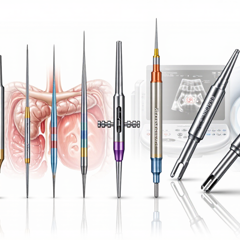
What Biopsy Needles Are and How They Support Accurate Tissue Sampling
Biopsy needles are slender medical instruments designed to collect cells or tissue for laboratory examination. They help clinicians investigate suspicious findings while limiting unnecessary tissue damage. Fine-needle aspiration uses a narrow needle to remove individual cells or fluid. Core biopsy needles obtain small tissue cylinders, preserving internal structure for more detailed analysis. The choice depends on the lesion, its location, and the diagnostic question.
Accurate sampling requires more than needle sharpness. Imaging guidance, such as ultrasound or computed tomography, helps the clinician place the needle precisely. Needle gauge, tip design, depth control, and a stabilizing sheath can influence specimen quality. A core needle may be preferred when pathologists need tissue architecture. Several carefully planned samples may be necessary, especially when a lesion contains mixed or uneven areas.
Patient safety and specimen handling matter just as much. Clinicians assess bleeding risk, nearby organs, prior imaging, and the safest access route before the procedure. The collected material should be labeled immediately and transferred under appropriate conditions. No needle is perfect. Even a technically successful pass can produce too little or poorly preserved tissue. Reviewing imaging findings with the pathology team can reduce that risk, although decisions still require clinical judgment. Sometimes, the most accurate plan changes during the procedure.
7 Best Biopsy Needles to Achieve Accurate Sampling - What Biopsy Needles Are and How They Support Accurate Tissue Sampling
| No. | Biopsy Needle Type | Typical Gauge | Sampling Mechanism | Specimen Obtained | Common Uses | How It Supports Accurate Sampling | Key Limitations |
|---|---|---|---|---|---|---|---|
| 1 | Fine-Needle Aspiration Needle | 22–25 G | Negative pressure draws cells and fluid through a narrow needle. | Cellular material, fluid, and cytology smears. | Thyroid, lymph nodes, superficial masses, and cystic lesions. | Small diameter can reduce tissue trauma and enables precise targeting under ultrasound or endoscopic guidance. | Usually provides limited tissue architecture and may yield insufficient material for some ancillary tests. |
| 2 | Core Cutting Needle | 14–20 G | A cutting cannula advances around a notched stylet to remove a tissue core. | Cylindrical tissue core with preserved architecture. | Liver, kidney, breast, prostate, soft tissue, and other solid-organ lesions. | Preserved tissue structure helps histopathology, grading, receptor testing, and assessment of lesion heterogeneity. | Larger needles may cause more bleeding or discomfort and require careful image-guided placement. |
| 3 | Automatic Spring-Loaded Core Needle | 14–20 G | A spring-driven mechanism rapidly advances the stylet and cutting cannula. | Multiple consistent core specimens. | Image-guided sampling of solid masses and parenchymal organs. | Rapid, reproducible firing can reduce hand motion and help produce uniform cores when the target is stable. | The firing action can cause needle movement; adequate safety margins and stable imaging guidance are essential. |
| 4 | Semi-Automatic Core Needle | 14–20 G | The operator manually advances the stylet, followed by spring-assisted deployment of the cutting cannula. | Core tissue specimens with preserved architecture. | Lesions close to critical structures and procedures requiring controlled needle positioning. | Manual stylet positioning allows the operator to control the sampling depth before the cutting action. | Typically requires two coordinated steps and may be slower than fully automatic systems. |
| 5 | Coaxial Biopsy System | Introducer: 13–19 G Inner needle: 14–20 G |
An outer introducer remains in position while the inner aspiration or core needle is exchanged through it. | One or more aspiration samples or tissue cores. | Lung, liver, kidney, bone, and deep lesions requiring repeat passes. | Maintains a stable access route, reduces repeated punctures, and facilitates multiple samples from slightly different target regions. | Adds device diameter and procedural complexity; it is an access system rather than a standalone sampling design. |
| 6 | Multi-Plane Franseen-Tip Needle | 19–22 G | Multiple cutting surfaces at the distal tip capture tissue during controlled advancement or rotation. | Histology-quality core fragments, often in a compact sample. | Endoscopic ultrasound-guided sampling of pancreatic, biliary, lymph-node, and other deep lesions. | The multi-edged tip can improve tissue capture and provide material suitable for architecture-dependent diagnosis. | Performance depends on lesion firmness, needle handling, imaging guidance, and the number of passes. |
| 7 | Menghini-Type Aspiration Needle | 16–22 G | A sharp bevel and suction are used to obtain a narrow tissue fragment. | Small core-like fragments and cellular material. | Selected liver and other soft-tissue sampling procedures, depending on local technique. | Combines aspiration with a cutting action and can obtain more tissue than conventional cytology-only aspiration. | Specimen size and quality can vary with suction, operator technique, tissue consistency, and needle positioning. |
Note: Gauge ranges are typical clinical ranges and may vary by procedure, anatomy, imaging method, and needle design. Needle selection should be based on the target lesion, required specimen type, bleeding risk, and operator experience.
Related Posts
-
Top 10 Coaxial Needle Suppliers in China You Should Know?
-
How to Choose the Right Bone Biopsy Needle for Your Medical Needs
-
China Top Tru Cut Biopsy Needle Manufacturers Suppliers
-

Discover the Best Argon Suppliers in Athens TX Today
-
2026 Best Arrow Arterial Line Kit for Efficient Medical Use?
-

How to Choose the Right Bone Marrow Biopsy Needle for Effective Results